
SEPTIC ARTHRITIS

Introduction: 

Septic arthritis is a medical emergency. It usually arises from haematogenous spread of bacterial infection from another site, commonly the skin or upper respiratory tract. Infection from direct puncture wounds or that secondary to joint aspiration is uncommon. Risk factors for septic arthritis include increasing age, pre-existing joint disease (especially RA), diabetes mellitus, immunosuppression and IV drug misuse.
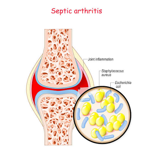


Clinical features

The usual presentation is with acute or subacute monoarthritis. The joint is usually swollen, hot and red, with pain at rest and on movement. The knee and hip are the most common sites. The usual culprit organism is Staphylococcus aureus. Disseminated gonococcal infection is another cause in young, sexually active adults. This presents with migratory arthralgia and low-grade fever, followed by the development of oligo- or monoarthritis. Painful pustular skin lesions may also be present. Lyme disease and brucellosis are less common causes of septic arthritis.


Investigations

Joint fluid aspiration for Gram stain and culture is essential, under image guidance if deep. Aspirated fluid often looks turbid or bloodstained.

Blood cultures may be positive due to bacteraemia. Blood tests may reveal leucocytosis with raised ESR and CRP, although these may be absent in elderly or immunocompromised patients.

Concurrent cultures from the genital tract are indicated if gonococcal infection is likely.


Management

Pain relief.

IV antibiotics: IV flucloxacillin 2 g three times daily is first choice until identification of the organism and its sensitivities is possible. IV treatment is usually continued for 2 wks, followed by oral treatment for a further 4 wks.

 Daily joint aspiration in the initial stages to minimise the effusion. If this is unsuccessful, surgical drainage may be required.

Early mobilisation.


Comments

Popular posts from this blog

BRONCHIECTASIS

INTRODUCTION: Bronchiectasis means abnormal dilatation of the bronchi due to chronic airway inflammation and infection. It is usually acquired, but may result from an underlying genetic or congenital defect of airway defences. CAUSES: Congenital • Cystic fibrosis • Primary ciliary dyskinesia • Kartagener’s syndrome (sinusitis and transposition of the viscera) • Primary hypogammaglobulinaemia Acquired • Pneumonia (complicating whooping cough or measles) • Inhaled foreign body • Suppurative pneumonia • Pulmonary TB • Allergic bronchopulmonary aspergillosis complicating asthma • Bronchial tumours CLINICAL FEATURES: ● Chronic cough productive of purulent sputum.  ● Pleuritic pain. ● Haemoptysis.  ● Halitosis. Acute exacerbations may cause fever and increase these symptoms. Examination reveals coarse crackles caused by sputum in bronchiectatic spaces. Diminished breath sounds may indicate lobar collapse. Bronchial breathing due to scarring may be heard in advanced disease. INVESTIG...

CARDIAC REHABILITATION

  Introduction “Cardiac Rehabilitation is the process by which patients with cardiac disease, in partnership with a multidisciplinary team of health professionals are encouraged to support and achieve and maintain optimal physical and psychosocial health. The involvement of partners, other family members and carers is also important”. Cardiac rehabilitation is an accepted form of management for people with cardiac disease. Initially, rehabilitation was offered mainly to people recovering from a myocardial infraction (MI), but now encompasses a wide range of cardiac problems. To achieve the goals of cardiac rehabilitation a multidisciplinary team approach is required. The multidisciplinary team members include: Cardiologist/Physician and co-coordinator to lead cardiac rehabilitation Clinical Nurse Specialist Physiotherapist Clinical nutritionist/Dietitian Occupational Therapist Pharmacist Psychologist Smoking cessation counsellor/nurse Social worker Vocational counsellor Clerical Ad...

லம்பார் ஸ்பாண்டிலோஸிஸ்(lumbar spondylosis)

  முன்னுரை ல ம்பார் ஸ்பாண்டிலோஸிஸ்(lumbar spondylosis) எனப்படும் மருத்துவ பிரச்சினைகள் என்பது முதுகுப் பகுதியில் ஏற்படும் நீண்ட நாள் முதுகு வலி. இவ்வாறு ஏற்படும் முதுகு வலி முதுகு முள்ளெலும்பு பகுதியில்(vertebral coloum) உள்ள தட்டு அழுத்தப் படுவதினால்(disk compression) அல்லது முள்ளெலும்பு பகுதியின பிரதான பகுதி சற்று இடம் நகர்வதால(displacement) முதுகு வலி ஏற்படுவதற்கு வாய்ப்புகள் உள்ளதாக கூறப்படுகின்றன. சில சமயங்களில் முதுகு தண்டு மற்றும் எலும்பு பகுதிகள் தொடர்சிதைவு(degeneration) ஆகும் போதும், முதுகெலும்பு தட்டு பகுதி, முதுகெலும்பு மூட்டு(facet joints) பகுதி தொடர்ந்து பிரச்சனைக்கு உள்ளாக்கப்படும் பொழுதும் முதுகு வலி ஏற்படுகிறது. ஸ்பாணடிலோஸிஸ் என்பதை முதுகு எலும்பு தேய்மானம்(osteoarthritis)  எ ன்று கூறலாம். இவ்வாறு முதுகு எலும்பு தேய்மானம்,  ல ம்பார்(lumbar vertebrae)  எனப்படும் கீழ் முதுகு எலும்பு பகுதிகள், மேல் முதுகு எலும்பு பகுதிகள்(thoracic vertebrae), மற்றும் கழுத்து முதுகெலும்பு(cervical vertebrae) பகுதிகள் போன்றவற்றை பாதிக்கலாம். பொதுவாக ஸ்பாண்டிலோசிஸ் எனப்படு...

CARDIAC ARREST AND RESUSCITATION

INTRODUCTION: The leading causes of sudden death before old age, in people over the age of 44, are ventricular fibrillation from asymptomatic ischaemic heart disease or non-traumatic accidents such as drowning and poisoning. In people under the age of 38, the commonest causes are traumatic, due to accident or violence. In such instances death may be prevented if airway obstruction can be reversed, apnoea or hypoventilation avoided, blood loss prevented or corrected and the person not allowed to be pulseless or hypoxic for more than 2 or 3 minutes. If, however, there is circulatory arrest for more than a few minutes, or if blood loss or severe hypoxia remain uncorrected, irreversible brain damage may result. Immediate resuscitation is capable of preventing death and brain damage. The techniques required may be used anywhere, with or without equipment, and by anyone, from the lay public to medical specialists, provided they have been appropriately trained. Resuscitation may be divided in...

PARKINSON'S DISEASE

  Parkinson's EtiologyParkinson's disease (PD) is a neurodegenerative disorder that mostly presents in later life with generalized slowing of movements (bradykinesia) and at least one other symptom of resting tremor or rigidity. Other associated features are a loss of smell, sleep dysfunction, mood disorders, excess salivation, constipation, and excessive periodic limb movements in sleep (REM behavior disorder). PD is a disorder of the basal ganglia, which is composed of many other nuclei. The striatum receives excitatory and inhibitory input from several parts of the cortex. The key pathology is the loss of dopaminergic neurons that lead to the symptom .  It is the seconds most common neuro-degenerative condition in the world after Alzheimer's. The condition is caused by the slow deterioration of the nerve cells in the brain, which create dopamine. Dopamine is a natural substance found in the brain that plays a major role in our brains and bodies by messag...

முதுகு வலி மற்றும் முதுகு தண்டுவட வலி உள்ளவர்களுக்கு கடைபிடிக்க வேண்டிய சில வழிமுறைகள்....

    முதுகு வலி மற்றும் முதுகு தண்டுவட வலி உள்ளவர்களுக்கு கடைபிடிக்க வேண்டிய சில வழிமுறைகள் ....     பொதுவாக முதுகு வலி என்பது இன்றைய காலகட்டத்தில் பல பேருக்கு மிக அதிகமாகவே காணப்படுகிறது. இவ்வாறு வலி இருக்கும் பொழுது என்ன மாதிரியான வழிமுறைகளை கடைப்பிடிக்க வேண்டும் என்பதை கீழே விரிவாக பார்க்கலாம்.   பொதுவாக முதுகு வலி ஆரம்பிக்கும் பொழுது அவற்றை உதாசீனப்படுத்தாமல் அருகில் உள்ள மருத்துவரை அணுகி ஆலோசனை பெறுவது மிகவும் முக்கியம். மேலும் முதுகு வலி ஏற்படும் பொழுது அவற்றுக்கு தேவையான மருத்துவம்(medical management), இயன்முறை மருத்துவம்(physiotherapy treatment), பயிற்சிகள்(exercises) அல்லது அறுவை சிகிச்சை(surgery) மற்றும் புனர்வாழ்வு சிகிச்சைகள்(Rehabilitation) போன்றவை தேவைப்படலாம். மேலே கண்ட மருத்துவத்தில் ஏதாவது ஒன்றை எடுத்துக் கொள்ளும் பட்சத்தில் மேலும் முதுகு வலி வராமல் பாதுகாத்துக் கொள்ளவும், நமது அன்றாட வேலைகளை தொடர்ந்து செய்யவும், மருத்துவ உபகரணங்களை பயன்படுத்திக் கொ...

RELAXED POSITIONS FOR BREATHLESS PATIENTS

Relaxation positions for the breathless patient  If patients can be taught how to control their breathing during an attack of dyspnoea, this can be of great benefit to them. The patient should be put into a relaxed position, and encouraged to do ‘diaphragmatic’ breathing at his own rate. The rate of breathing does not matter at this stage; it is the pattern of breathing that is important. As the patient gains control of his breathing he should be encouraged to slow down his respiratory rate. Any of the following positions will assist relaxation of the upper chest while encouraging controlled diaphragmatic breathing. They can be adapted to various situations in everyday life. HIGH SIDE LYING  Five or six pillows are used to raise the patient’s shoulders while lying on his side. One pillow should be placed between the waist and axilla, to keep the spine straight and prevent slipping down the bed. The top pillow must be above the shoulders, so that only the head and neck are supp...